Home

Solve dam Installation cross section of retina Contributor Billy goat Weave

View Image
View Image

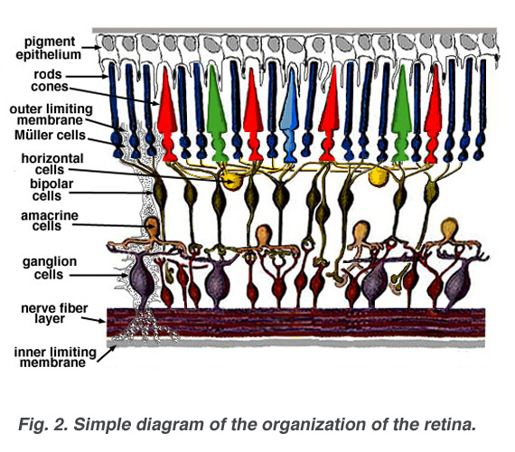
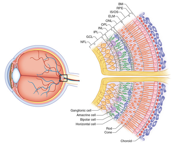
Simple Anatomy of the Retina by Helga Kolb – Webvision

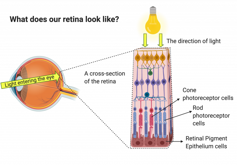
The retina and retinal pigment epithelium (RPE) | UCL Institute of  Ophthalmology - UCL – University College London
The retina and retinal pigment epithelium (RPE) | UCL Institute of Ophthalmology - UCL – University College London
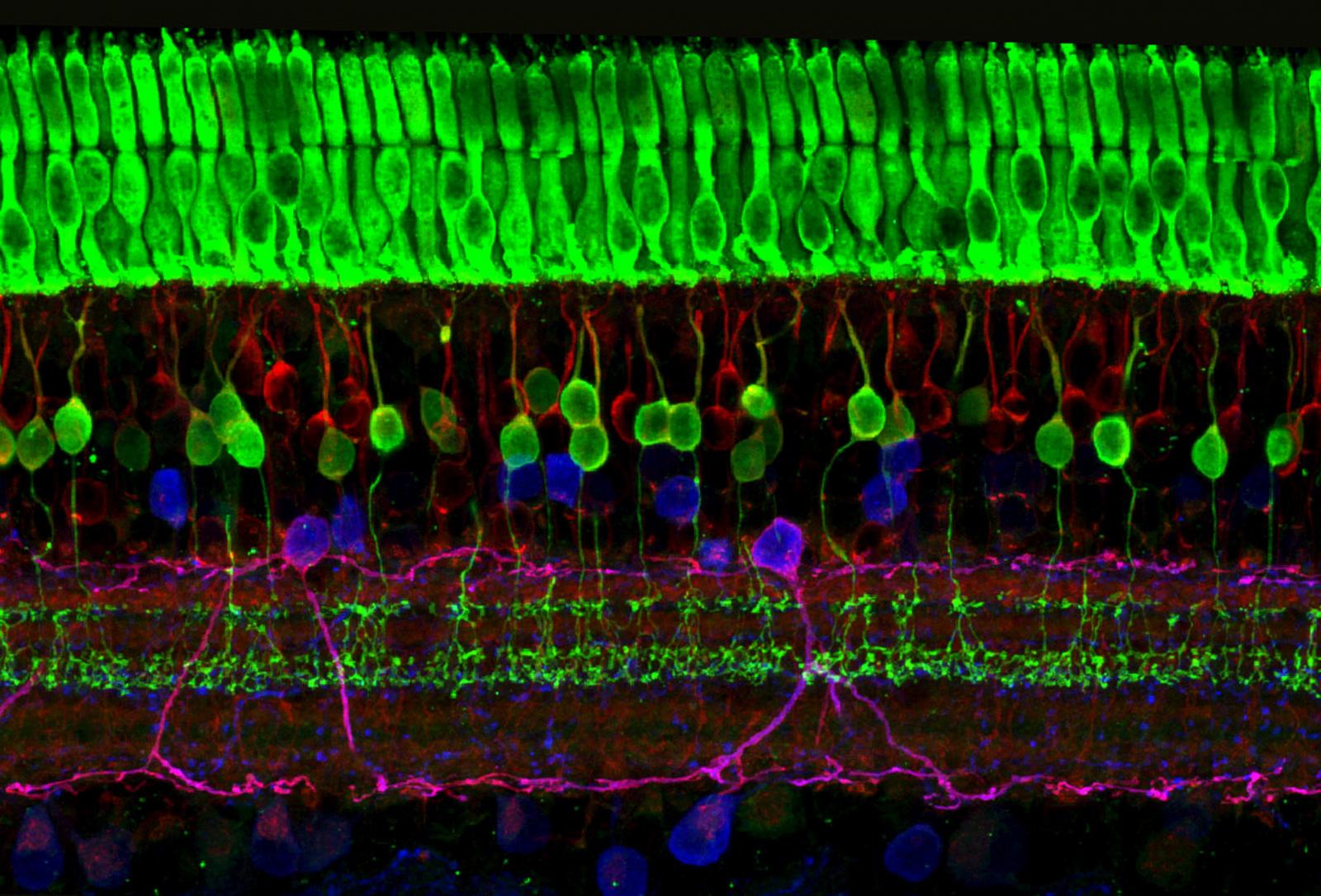
neuroscience - How do cone cells underneath capillaries receive light? -  Biology Stack Exchange
neuroscience - How do cone cells underneath capillaries receive light? - Biology Stack Exchange

Figure 12. [Schematic of a retinal cross-section...]. - Webvision - NCBI  Bookshelf
Figure 12. [Schematic of a retinal cross-section...]. - Webvision - NCBI Bookshelf

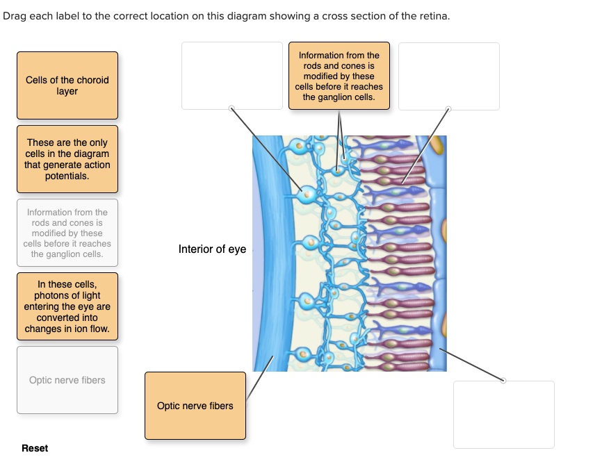
SOLVED: Drag each label to the correct location on this diagram showing cross  section of the retina. Information from the rods and cones is modified by  these cells before reaches the ganglion
SOLVED: Drag each label to the correct location on this diagram showing cross section of the retina. Information from the rods and cones is modified by these cells before reaches the ganglion
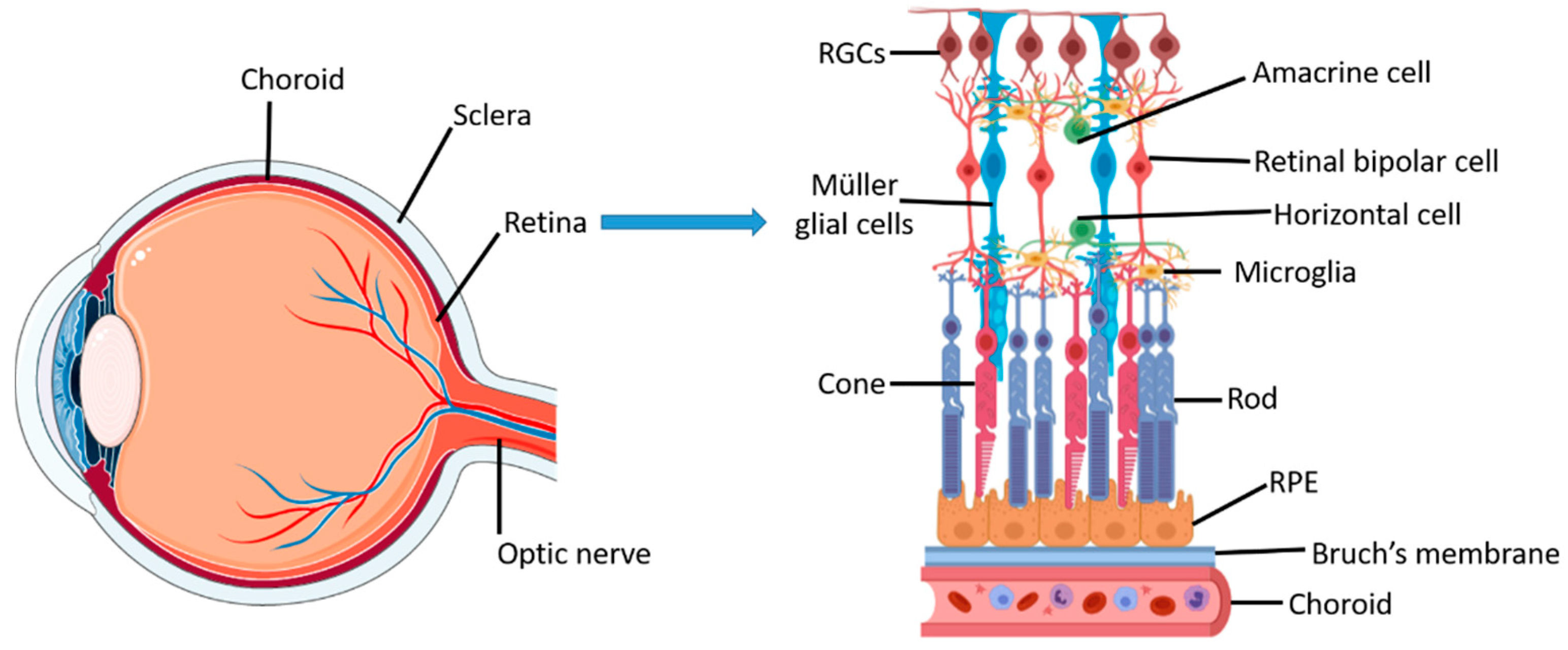
File:Human eye cross section detached retina.svg - Wikimedia Commons

Applied Sciences | Free Full-Text | The Evolution of Fabrication Methods in  Human Retina Regeneration
Applied Sciences | Free Full-Text | The Evolution of Fabrication Methods in Human Retina Regeneration

Parts of the Eye
Parts of the Eye

2020–2021 BCSC Basic and Clinical Science Course™

The Chemistry of Human Vision – The Retinoid Cycle

Anatomy of the Eye - Retina-Vitreous Surgeons of CNY

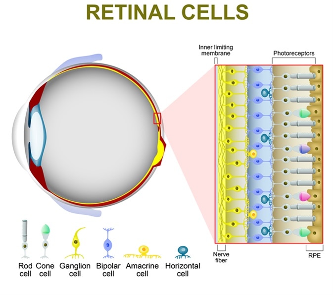
Eye Anatomy. Rod Cells And Cone Cells. The Arrangement Of Retinal Cells Is  Shown In A Cross Section. Vector Diagram For Your Design, Educational,  Biological, Science And Medical Use Royalty Free SVG,
Eye Anatomy. Rod Cells And Cone Cells. The Arrangement Of Retinal Cells Is Shown In A Cross Section. Vector Diagram For Your Design, Educational, Biological, Science And Medical Use Royalty Free SVG,

13.1A: Anatomy of the Eye - Medicine LibreTexts

Schematic cross-section showing the retinal blood vessels lining the... |  Download Scientific Diagram
Schematic cross-section showing the retinal blood vessels lining the... | Download Scientific Diagram
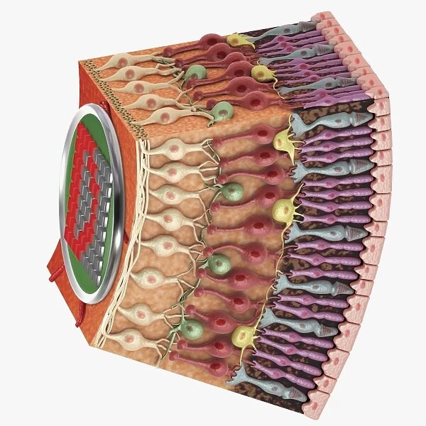
Cross section digital illustration of retina with available as Framed  Prints, Photos, Wall Art and Photo Gifts #13546487
Cross section digital illustration of retina with available as Framed Prints, Photos, Wall Art and Photo Gifts #13546487
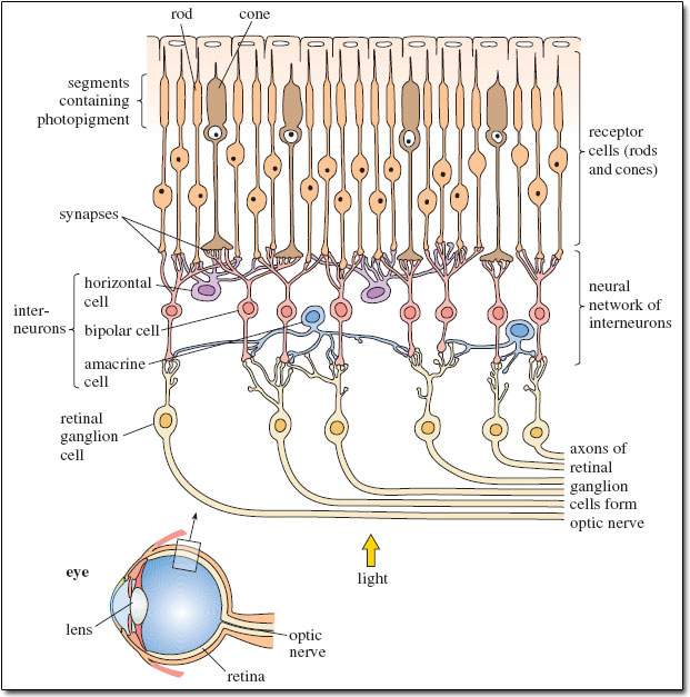
Figure 6. [Cross-section of the human retina...]. - Webvision - NCBI  Bookshelf
Figure 6. [Cross-section of the human retina...]. - Webvision - NCBI Bookshelf

Retinal Neurophysiology Section | National Eye Institute

Simple Anatomy of the Retina by Helga Kolb – Webvision

Studying mammals: The social climbers: Figure 2 Schematic cross-section of  the retina. If you look at the arrow labelled 'light' and the small inset  diagram of the structure of the eye, you'll
Studying mammals: The social climbers: Figure 2 Schematic cross-section of the retina. If you look at the arrow labelled 'light' and the small inset diagram of the structure of the eye, you'll

JCI - Therapeutic targets in age-related macular disease

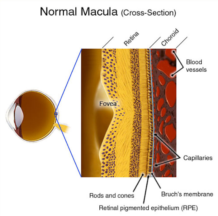
Normal Retinal Anatomy - The Retina Reference

How the Body Works : Cross Section of an Eyeball - YouTube

Cross-section schematic of the eye with enlargement of the retina. |  Download Scientific Diagram
Cross-section schematic of the eye with enlargement of the retina. | Download Scientific Diagram